Réunion
Journée thématique Imagerie vasculaire 2026
Axes scientifiques :
- Apprentissage machine
- Audio, Vision et Perception
Organisateurs :
Nous vous rappelons que, afin de garantir l'accès de tous les inscrits aux salles de réunion, l'inscription aux réunions est gratuite mais obligatoire.
Inscriptions
26 personnes membres du GdR IASIS, et 30 personnes non membres du GdR, sont inscrits à cette réunion.
Capacité de la salle : 140 personnes. 84 Places restantes
Inscriptions closes pour cette journée
Annonce
Nous proposons cette deuxième édition avec la volonté de soutenir les actions pluridisciplinaires à l’interface entre les activités de recherche du GDR (apprentissage artificiel, traitement du signal, vision artificielle) et les problématiques cliniques en lien avec l’imagerie vasculaire. De plus, nous souhaitons mettre en valeur la valorisation industrielle, les développements reproductibles et open-source pouvant bénéficier au plus grand nombre dans les communautés scientifiques et médicales.
Les thèmes abordés sont, sans être exhaustif :
- Imagerie médicale, radiologie vasculaire, cardiaque, hépatique, etc.
- Thérapies guidées par l’image, technologies médicales
- Analyse et traitement d’images médicales et biomédicales
- Apprentissage artificiel pour l’imagerie médicale et la simulation
- Développement logiciel open-source
- Simulation et modélisation numériques de mécanismes biologiques
- Annotation automatique ou assistée de données médicales
Cette journée pourrait donc concerner des chercheurs en traitement du signal, en informatique, en vision artificielle, en mathématiques appliquées, etc. et des médecins en radiologie vasculaire ou en chirurgie vasculaire par exemple.
Comité d’organisation
- Antoine Vacavant, Institut Pascal, Université Clermont Auvergne, antoine.vacavant@uca.fr
- Bertrand Kerautret, LIRIS, Université Lumière Lyon 2, bertrand.kerautret@univ-lyon2.fr
- Nicolas Passat, CReSTIC, Université de Reims Champagne Ardenne, nicolas.passat@univ-reims.fr
Programme de la journée
Le planning complet est le suivant :
| 08h30-09h00 | Accueil des participants |
| 09h00-09h15 | Mot d’accueil |
| 09h15-10h00 | Conférence invitée scientifique – Marcela SZOPOS Combining Physics-Based Modeling and Data Analytics Methods To Develop a Digital Twin for Ocular Applications |
| 10h00-10h15 | Fast track – Présentations courtes des posters |
| 10h15-10h45 | Pause café & session posters/démos |
| 10h45-12h00 | Présentations sur sélection – Session IA & Imagerie |
| 12h00-13h45 | Pause déjeuner |
| 13h45-14h30 | Conférence invitée médicale – Guillaume CANAUD From a single patient to a US FDA approved drug: the story of a drug repositioning for a rare disease |
| 14h30-15h30 | Présentations sur sélection – Session Recherche Translationnelle |
| 15h30-16h00 | Pause café & session posters/démos |
| 16h00-16h45 | Conférence invitée industrielle – Laurence ROUET Vascular image quantification |
| 16h45-17h30 | Présentations sur sélection – Session Traitement du Signal |
| 17h30-18h00 | Fin de la journée |
Programme détaillé
- 09h15-10h00 – Conférence scientifique : Marcela SZOPOS – MAP5 / Univ. Paris Cité
Titre : Combining Physics-Based Modeling and Data Analytics Methods To Develop a Digital Twin for Ocular Applications
Résumé : Understanding the behavior of the human eye is challenging due to the complex interactions between various physical phenomena, such as fluid dynamics, the deformation of surrounding tissues and the impact of their temperature. Although medical data can offer valuable insights into ocular physiopathology, the available information can be scarce and noisy. Moreover, in experimental studies, multiple factors come into play and it is difficult to isolate the contributions of individual mechanisms. Therefore, developing a robust and accurate digital twin for ocular applications can enhance our understanding of this complex system, by integrating governing mechanisms and data variability.
This talk presents our ongoing efforts in this direction. First, we focus on assessing the combined effects of ocular blood flow and different ocular tissues by means of a coupled multiscale hemodynamics and biomechanics model [1]. Secondly, building upon previous developments in retinal blood flow modeling, we integrate clinical measurements and physiological insights with the aim of providing a robust, clinically relevant analysis of waveform parameters in the retinal vasculature [2]. Finally, we highlight the potential of the model for studying cardiovascular diseases with ocular manifestations [3]. We will illustrate these developments, discuss their applications in ocular physiology and treatment, and address remaining challenges, including inverse problems and parameter identification.
[1] L. Sala et al. The ocular mathematical virtual simulator: A validated multiscale model for hemodynamics and biomechanics in the human eye, Int. J. Numer. Meth. Biomed. Engng. (2024).
[2] L. Sala et al. Analysis of waveform parameters in the retinal vasculature via mathematical modeling and data analytics methods, La Matematica (2024).
[3] L. Sala et al. Eye2Heart: A reduced mathematical model bridging cardiovascular and ocular hemodynamics, Math. Biosci. Engng (2026). - 10h00-10h15 – Présentations posters & démos
– Arthur LOUVART, Kahina BENSAFIA-CHERFA, Wit HAERTLE, Ali MANSOUR, Clément HOFFMANN : SEGMENTATION PAR IA ET ESTIMATION AUTOMATIQUE DE LA SURFACE DES PLAIES VASCULAIRES
– Luc ANCHLING, Adrian BASARAB, Nicolas MELLADO, Fabien VIDAL : VESSEL PUNCTURE USING ULTRASOUND-FREE NEEDLE GUIDANCE BASED ON AUGMENTED REALITY
– Damien RON, Ningyuan ZHANG, Marine CAMUS, Isabelle CLAUDE : RECONSTRUCTION 3D AVANCEE DES VOIES BILIAIRES ET DU FOIE POUR LA CHOLANGIOPANCREATOGRAPHIE RETROGRADE PAR VOIE ENDOSCOPIQUE (CPRE)
– Manel MEFTAH, Benoît NAEGEL, Adrien KRAHENBUHL: SQUELETTISATION D’OBJETS TUBULAIRES PAR ACCUMULATION DISCRETE DE NORMALES - 10h45-12h00 – Session IA & Imagerie
– Nathan PAINCHAUD, Tristan HABEMONT, Morgane DES LIGNERIS, Allan SERVA, Pierre CROISILLE, Laurent BERTOLETTI, Thomas LAMPERT, Johannes F. LUTZEYER, Odyssée MERVEILLE : PULMONARY EMBOLISM RISK STRATIFICATION FROM CTPA AND MEDICAL RECORDS: VASCULAR GRAPHS ARE NOT ALL YOU NEED
– Daniele FALCETTA, Liane S. CANAS, L. SUPPA, M. PENTASSUGLIA, X. ZHANG, J. CLEARY, M. MODAT, S. OURSELIN, Maria A. ZULUAGA : CARAVELMETRICS : UN CADRE AUTOMATISE POUR L’ANALYSE CEREBROVASCULAIRE A GRANDE ECHELLE BASEE SUR LES GRAPHES
– Da CHEN, Laurent D. COHEN : CURVATURE-PENALIZED GEODESIC MODELS FOR VASCULAR CENTERLINE EXTRACTION
– Halima FOUADI, Mohamed KAS, Yassine RUICHEK, Youssef EL MERABET : SYNTHESE ET SEGMENTATION CARDIAQUES CONTROLEES PAR LE TEXTE : DE LA GENERATION D’IRM 2D A LA MODELISATION VOLUMETRIQUE 3D
– Abir AISSAA, Florence ROSSANT, Hélène URIEN, Michel PAQUES : PIPELINE D’ANALYSE AUTOMATISEE D’IMAGES EN OPTIQUE ADAPTATIVE POUR L’ETUDE DE LA VASCULARISATION RETINIENNE - 13h45-14h30 – Conférence médicale : Guillaume CANAUD – Institut Necker-Enfants malades
Titre : From a single patient to a US FDA approved drug: the story of a drug repositioning for a rare disease
Résumé : CLOVES syndrome (congenital lipomatous overgrowth, vascular malformations, epidermal naevi, scoliosis/skeletal and spinal syndrome) is a genetic disorder that results from somatic, mosaic gain-of-function mutations of the PIK3CA gene, and belongs to the spectrum of PIK3CA-related overgrowth syndromes (PROS). This rare condition has no specific treatment and a poor survival rate. Here, we describe a postnatal mouse model of PROS/CLOVES that partially recapitulates the human disease, and demonstrate the efficacy of BYL719, an inhibitor of PIK3CA, in preventing and improving organ dysfunction. On the basis of these results, we used BYL719 to treat nineteen patients with PROS. The drug improved the disease symptoms in all patients. Previously intractable vascular tumours became smaller, congestive heart failure was improved, hemihypertrophy was reduced, and scoliosis was attenuated. The treatment was not associated with any substantial side effects. In conclusion, this study provides the first direct evidence supporting PIK3CA inhibition as a promising therapeutic strategy in patients with PROS. - 14h30-15h30 – Session Recherche Translationnelle
– Raoul SALLÉ DE CHOU, Laurent NAJMAN, Hugues TALBOT, Irène VIGNONCLEMENTEL : SIMULATION DE PERFUSION DU MYOCARDE REPOSANT SUR DES METHODES D’APPRENTISSAGE AUTOMATIQUE
– Dennis-Jose DA-SILVA, Alexandra FONTAINE, Laurent DEBELLE, Nicolas PASSAT, Sébastien ALMAGRO : ETUDE DES ALTERATIONS AORTIQUES RELIEES AU DIABETE PAR TOMOGRAPHIE X SYNCHROTRON
– Mathilde DUPOUY, Yamil VINDAS, Thibaut DAMBRY, Blaise Kévin GUEPIE, Philippe DELACHARTE : CLASSIFICATION OF CEREBRAL MICROEMBOLI : LATENT SPACE ANALYSIS AND UNCERTAINTY QUANTIFICATION FOR SOFTWARE INTEGRATION INTO A TRANSCRANIAL DOPPLER MEDICAL DEVICE
– René ANXIONNAT, Marine BEAUMONT, Bailiang CHEN, Tae-Hee CHO, Mégane DECROOCQ, Morgane DES LIGNERIS, Guillaume DOLLE, Esther FONTAINE, Carole FRINDEL, Benjamin GORY, François-Régis HAMMER, Radhouane JILANI, Erwan KERRIEN, Antoine KNEIB, Odyssée MERVEILLE, Émilien MICARD, Nicolas PASSAT, Christophe PORTEFAIX, Ulysse PUEL, Pierre ROUGE, Stéphanie SALMON, Pierre-Frédéric VILLARD, François ZHU : SIMULATION PREDICTIVE POUR LA PLANIFICATION EN NEURORADIOLOGIE INTERVENTIONNELLE : BILAN D’UN PROJET COLLABORATIF - 16h00-16h45 – Conférence industrielle : Laurence ROUET – SIEMENS Healthineers
Titre : Vascular image quantification
Résumé : The presentation will focus on two arterial territories associated with different pathological conditions. In a first part, we will present atherosclerosis and the imaging modalities used for patient monitoring. Then we will explain how carotid plaque quantification is associated to cardio-vascular events and present the successive steps that enabled to build an AI model of plaque. In a second part, we will present an approach for the quantification of abdominal aortic strain in the context of better assessment of the risk of wall rupture. We will present the required imaging acquisitions, their processing and how the results can contribute to better tissue characterization.
Finally, we will open research perspectives related to the new advanced vascular imaging lab that we are setting up with INSERM and CHU Nantes, in a public-private partnership with Siemens Healthineers. - 16h45-17h30 – Session Traitement du Signal
– Dina ABDALLAH, Barbara NICOLAS, Ewen CARCREFF, Hervé LIEBGOTT : FEASIBILITY OF 3D CLUTTER-FILTERED WAVE IMAGING FOR MECHANICAL WAVE TRACKING IN ARTERIAL WALLS– Franck PLOURABOUE : REVEALING THE VASCULAR SIGNATURE OF INTRA-CRANIAL PRESSURE
– Adrien GORDON, Xinmiao QIU, Grégoire BUISSON, Chloé PAQUET, Yann FISCHER, Zacharie AURAY, Marius DUBOSC, Jules GUILLOU, Maxime Boy ARNOULD, Michael ATLAN: QUANTITATIVE MICROVASCULAR HEALTH ENDPOINTS FROM RETINAL LASER DOPPLER HOLOGRAPHY
Soumettre un résumé
Vous souhaitez présenter vos travaux de recherche ou de valorisation en imagerie vasculaire ? Envoyez aux membres du Comité d’organisation un résumé en suivant le template fourni ici, par email, et avant la date indiquée dans la section Dates importantes.
Dates importantes
| 15 avril 2026 | Date limite d’envoi d’un résumé |
| 17 avril 2026 | Sélection des résumés et mise en place du programme |
| 17 avril 2026 | Diffusion du programme final de la journée |
| 11 mai 2026 | Journée thématique |
Résumés des contributions
Marcela SZOPOS - MAP5 / Univ. Paris Cité
Titre : Combining Physics-Based Modeling and Data Analytics Methods To Develop a Digital Twin for Ocular Applications
Résumé : Understanding the behavior of the human eye is challenging due to the complex interactions between various physical phenomena, such as fluid dynamics, the deformation of surrounding tissues and the impact of their temperature. Although medical data can offer valuable insights into ocular physiopathology, the available information can be scarce and noisy. Moreover, in experimental studies, multiple factors come into play and it is difficult to isolate the contributions of individual mechanisms. Therefore, developing a robust and accurate digital twin for ocular applications can enhance our understanding of this complex system, by integrating governing mechanisms and data variability.
This talk presents our ongoing efforts in this direction. First, we focus on assessing the combined effects of ocular blood flow and different ocular tissues by means of a coupled multiscale hemodynamics and biomechanics model [1]. Secondly, building upon previous developments in retinal blood flow modeling, we integrate clinical measurements and physiological insights with the aim of providing a robust, clinically relevant analysis of waveform parameters in the retinal vasculature [2]. Finally, we highlight the potential of the model for studying cardiovascular diseases with ocular manifestations [3]. We will illustrate these developments, discuss their applications in ocular physiology and treatment, and address remaining challenges, including inverse problems and parameter identification.
[1] L. Sala et al. The ocular mathematical virtual simulator: A validated multiscale model for hemodynamics and biomechanics in the human eye, Int. J. Numer. Meth. Biomed. Engng. (2024).
[2] L. Sala et al. Analysis of waveform parameters in the retinal vasculature via mathematical modeling and data analytics methods, La Matematica (2024).
[3] L. Sala et al. Eye2Heart: A reduced mathematical model bridging cardiovascular and ocular hemodynamics, Math. Biosci. Engng (2026).
Guillaume CANAUD - Institut Necker-Enfants malades
Titre : From a single patient to a US FDA approved drug: the story of a drug repositioning for a rare disease
Résumé : CLOVES syndrome (congenital lipomatous overgrowth, vascular malformations, epidermal naevi, scoliosis/skeletal and spinal syndrome) is a genetic disorder that results from somatic, mosaic gain-of-function mutations of the PIK3CA gene, and belongs to the spectrum of PIK3CA-related overgrowth syndromes (PROS). This rare condition has no specific treatment and a poor survival rate. Here, we describe a postnatal mouse model of PROS/CLOVES that partially recapitulates the human disease, and demonstrate the efficacy of BYL719, an inhibitor of PIK3CA, in preventing and improving organ dysfunction. On the basis of these results, we used BYL719 to treat nineteen patients with PROS. The drug improved the disease symptoms in all patients. Previously intractable vascular tumours became smaller, congestive heart failure was improved, hemihypertrophy was reduced, and scoliosis was attenuated. The treatment was not associated with any substantial side effects. In conclusion, this study provides the first direct evidence supporting PIK3CA inhibition as a promising therapeutic strategy in patients with PROS.
Laurence ROUET - SIEMENS Healthineers
Titre : Vascular image quantification
Résumé : The presentation will focus on two arterial territories associated with different pathological conditions. In a first part, we will present atherosclerosis and the imaging modalities used for patient monitoring. Then we will explain how carotid plaque quantification is associated to cardio-vascular events and present the successive steps that enabled to build an AI model of plaque. In a second part, we will present an approach for the quantification of abdominal aortic strain in the context of better assessment of the risk of wall rupture. We will present the required imaging acquisitions, their processing and how the results can contribute to better tissue characterization.
Finally, we will open research perspectives related to the new advanced vascular imaging lab that we are setting up with INSERM and CHU Nantes, in a public-private partnership with Siemens Healthineers.
-

![]() Expertises du GdR - cartographie par Axes - 19/09/2025
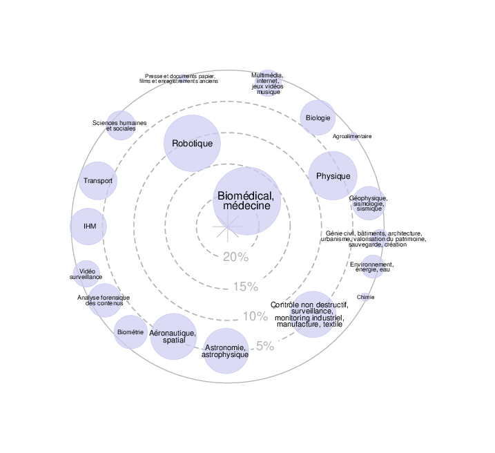
Expertises du GdR - cartographie par Axes - 19/09/2025![]() Expertises du GdR - cartographie par mots-clés applicatifs - 19/09/2025
Expertises du GdR - cartographie par mots-clés applicatifs - 19/09/2025