Réunion
Imagerie ultrasonore, de la formation d'images à leur analyse
Axes scientifiques :
- Imagerie computationnelle
Organisateurs :
Nous vous rappelons que, afin de garantir l'accès de tous les inscrits aux salles de réunion, l'inscription aux réunions est gratuite mais obligatoire.
Inscriptions
16 personnes membres du GdR IASIS, et 35 personnes non membres du GdR, sont inscrits à cette réunion.
Capacité de la salle : 50 personnes. -1 Places restantes
Inscriptions closes pour cette journée
Annonce
Attention au changement de lieu de la journée (voir ci-dessus).
NB. Déjeuner à la cantine de la délégation CNRS sera possible (paiement par CB).
Les images ultrasonores présentent des caractéristiques spécifiques (speckle important, résolution anisotrope, artefacts) qui les distinguent d’autres types d’imagerie et rendent nécessaire le développement de méthodes dédiées, aussi bien pour la reconstruction que pour l’analyse d’images.
Les avancées technologiques récentes dans les systèmes ultrasonores (électronique à grand nombre de voies, sondes matricielles) ouvrent de nouveaux défis méthodologiques, notamment pour l’imagerie 3D rapide. Il s’agira de repenser ou d’adapter les méthodes de traitement, mais également de réfléchir à leur accélération et à leur portabilité dans des applications cliniques ou industrielles.
Cette journée vise à rassembler les chercheurs et chercheuses s’intéressant à l’imagerie ultrasonore, suivant deux axes de recherche :
- formation d’images : stratégies d’acquisition, algorithmes d’imagerie, problèmes inverses, …
- analyse d’images : segmentation, recalage, ...
Bien qu’une majorité de la communauté scientifique en imagerie ultrasonore travaille sur des applications biomédicales, cette journée est ouverte à d’autres applications (comme le contrôle non-destructif industriel) qui en partagent certaines problématiques, contraintes physiques et méthodes.
La journée comportera 3 présentations invitées :
- Alexandre Aubry (Institut Langevin, ESPCI Paris)
- Diana Mateus (LS2N, Centrale Nantes)
- Samuel Rodriguez (I2M, Université de Bordeaux)
Appel à contributions
Nous invitons les jeunes chercheurs et chercheuses à soumettre une proposition pour présenter leurs travaux lors de l’événement (en anglais ou en français)
Les propositions sont attendues avant le 18 octobre, sous la forme d’un document comportant : titre, auteurs, affiliations, résumé de 15-20 lignes. Elles sont à envoyer par courriel à tous les organisateurs :
- Sébastien Bourguignon <sebastien.bourguignon@ec-nantes.fr>
- Clément Huneau <clement.huneau@univ-nantes.fr>
- Barbara Nicolas <barbara.nicolas@creatis.insa-lyon.fr>
- Valentine Wargnier-Dauchelle <valentine.wargnier@creatis.insa-lyon.fr>
Les notifications seront envoyées aux auteurs le 20 octobre.
Organisateur.ice.s
- Sébastien Bourguignon (LS2N, Centrale Nantes)
- Clément Huneau (LS2N, Nantes Université)
- Barbara Nicolas (CREATIS, CNRS)
- Valentine Wargnier-Dauchelle (CREATIS, INSA Lyon)
Programme
9h30-9h40 : Ouverture
9h40-10h20 : A. Aubry (invité), Institut Langevin, ESPCI Paris
10h20-10h45 : H. Mergui, LIP6 / Sonio SAS, Paris
10h45-11h10 : C. Galata, ICM, Paris
11h10-11h25 : Pause
11h25-11h50 : N. Moyne, Creatis, Lyon
11h50-12h15 : L. G. Pham, PhysMed, Paris
12h15-12h40 : S. Melin, LabTau, Lyon
12h40-14h00 : Pause Déjeuner
14h00-14h40 : S. Rodriguez (invité), I2M, Bordeaux
14h40-15h05 : J. E. Ayisah, LS2N / TPAC, Nantes
15h05-15h30 : Y. Sun, IMVIA, Dijon
15h30-15h45 : Pause
15h45-16h25 : D. Mateus (invitée), LS2N, Nantes
16h25-16h50 : D. Abdallah, Creatis, Lyon
16h50-17h15 : B. Hériard-Dubreuil, Inst. Langevin, Paris
Résumés des contributions
Présentations invitées
Ultrasound matrix imaging for joint wave-speed tomography and aberration correction in soft tissues
Alexandre Aubry, ESPCI Paris, Institut Langevin
Ultrasound imaging traditionally relies on the assumptions of single scattering and a constant speed of sound. In practice, spatial variations in sound speed within biological tissues distort propagating wave-fronts, severely degrading both spatial resolution and image contrast. To address these limitations, we present a physics-based computational framework for joint sound-speed tomography and aberration correction in soft tissues.
Experimentally, this approach leverages the acquisition of the reflection matrix, which captures the full set of impulse responses between the transducers of the ultrasonic probe. By decoupling the input and output focal points during the beamforming process, matrix imaging yields a focused reflection matrix that contains significantly richer information than conventional ultrasound images. Crucially, this representation enables local assessment of focusing quality to quantify wave aberrations at any point in the medium.
To recover the sound speed map, we propose a two-step strategy. First, inspired by astronomical adaptive optics, a multi-conjugate approach analyzes wavefront distortions across multiple depths. The mapped phase shifts optimally compensate for aberrations and allow a first reconstruction of the sound-speed spatial distribution. Second, wave propagation is modeled as a differentiable multi-layer network, leveraging optimization tools to solve the inverse problem and infer high-resolution sound-speed maps. We validate this framework through experiments on tissue-mimicking phantoms and human breast tissues, demonstrating its strong potential for tissue characterization and tumor detection.
Imagerie acoustique en milieu réverbérant : Localisation active de défauts et émission acoustique
Samuel Rodriguez, Université de Bordeaux, I2M
Les méthodes classiques d’auscultation de milieux solides ou biologiques par ondes acoustiques s’appuient sur une configuration expérimentale impliquant un trajet direct de l’onde entre la zone à inspecter et les transducteurs réalisant la mesure. Cela permet une exploitation facilitée des données mais prive de l’information portée par les autres interactions de l’onde avec le milieu. Le travail présenté ici s’attache à exploiter des champs acoustiques réverbérés, résultats de réflexions multiples sur les bords de la pièce et/ou de superpositions multiples dues à la géométrie. Pour l’inspection active de structures, le canevas méthodologique de traitement des données choisi est l’imagerie topologique qui permet de tirer avantage de cette réverbération notamment pour localiser des défauts qui ne sont pas en vue directe des transducteurs.
L’application industrielle associée actuellement développée au laboratoire est le contrôle des parties basses des rails de chemin de fer. Pour l’inspection passive de structures, les travaux se concentrent sur l’émission acoustique, une technique de suivi et de localisation des événements acoustiques générés dans et par la structure. L’utilisation du champ réverbéré plutôt que des ondes directs permet de réduire le nombre de transducteurs tout en conservant une résolution adaptée. Dans ce travail, nous mettons en œuvre de telles méthodes sur des réservoirs d’hydrogène à l’aide de transducteurs flexibles imprimés à façon. Le couplage de ces méthodes et de cette technologie est tourné vers le suivi de santé structurelle embarqué (SHM).
Implicit neural networks for representing ultrasound images and volumetric shapes
Diana Mateus, Centrale Nantes, LS2N
Neural Implicit Representations (INRs) have emerged as a versatile framework for medical imaging, providing continuous and compact representations that can effectively capture both image appearance and anatomical structure. Recent work has demonstrated their potential in ultrasound imaging, where they enable improved volumetric reconstruction, probe tracking, and raw data representation for applications such as novel view synthesis, compounding, and compression. Beyond image reconstruction, INRs have also proven effective as shape priors across populations, reducing annotation requirements for 3D segmentation and enabling unsupervised anomaly detection by modeling normal anatomical variability. The purpose of this talk is to show the versatility of INRs in ultrasound imaging.
Communications
Evaluating the realism of synthetic fetal cardiac ultrasound: speckle as the blind spot of generic metrics
Hanna Mergui, LIP6 / Sonio SAS, Paris
Synthetic images produced by diffusion models are increasingly used to compensate for the lack of annotated ultrasound data. Yet their evaluation mostly relies on generic metrics inherited from computer vision (FID, KID), which are largely insensitive to the specific properties of ultrasound images. We propose CARES, a five-phase evaluation protocol: anatomical fidelity, structural comparison, algorithmic utility, statistical and physical metrics, and expert perception. We applied it to 4,000 fetal echocardiograms generated by a latent diffusion model fine-tuned on 20,983 real images (four standard views, 60 sites). The evaluation axes disagree. The view least compliant with anatomical guidelines (14% of criteria met vs 67% for real images) was still recognized in 98.5% of cases by a clinical view-detection algorithm. It also gave the largest data-augmentation gain (+8.5 sensitivity points with 100 real images per view). The physical analysis reveals a bias specific to ultrasound. The signal-to-noise ratio is inflated 1.66- to 1.84-fold, with only 0.11–0.23 distribution overlap, whereas the contrast-to-noise ratio remains close to real data. The generator therefore reproduces tissue interfaces but smooths out speckle. Expert reading confirms overly homogeneous speckle and insufficient attenuation. Domain-adapted metrics, computed with a feature extractor trained on fetal ultrasound, widen the gap that ImageNet-based metrics underestimate. Finally, perceived realism varies widely across readers (fooling rates from 4.5% to 82%). These results call for evaluation criteria that account for ultrasound physics, and point to what must change for generative models to render speckle faithfully.
Patient-Specific Intraoperative Ultrasound Segmentation for Real-Time Trackerless Navigation in Brain Tumor Surgery
Caterina Galata, ICM, Paris
Intraoperative ultrasound (iUS) enables cost-effective, real-time visualization of residual tumor during brain tumor surgery, but remains difficult to interpret due to low tissue contrast, speckle, imaging artifacts, and inter-patient variability. Automatic segmentation of the tumor in iUS could enable trackerless navigation during surgery, yet existing deep-learning approaches are largely patient-agnostic and generalize poorly across patients and acquisition settings. We propose a patient-specific framework that trains a segmentation network exclusively on synthetic iUS generated from each patient’s preoperative data. The preoperative MRI volume and its tumor delineation are used to simulate iUS sweep trajectories, along which sequences of virtual 2D MRI and label slices are extracted within an ultrasound field of view. A pre-trained MRI-to-iUS synthesizer translates these slices into synthetic iUS images, which serve as training data for a lightweight 2D U-Net. The approach adapts segmentation to the patient’s anatomy and to the surgical target without requiring manually annotated iUS. We evaluate the framework on real iUS volumes from the ReMIND dataset. Tumor delineations were annotated independently on the MRI by two neurosurgeons (N1, n=10; N2, n=7) and transferred to iUS by 3D image registration. We compare our method with patient-agnostic U-Nets trained on public iUS datasets (RESECT-SEG and BraTioUS) and with an optical probe-tracking system (Brainlab Curve). Our trackerless method achieves the highest median Dice scores (80.8% on N1, 83.0% on N2), with median ASSD of 1.9 mm and 1.4 mm. Its Dice scores exceed those of the strongest patient-agnostic baseline by 18.4 and 23.8 percentage points, respectively, and are comparable to Brainlab Curve (78.0% and 79.0%), while requiring no specialized tracking hardware and running in real time (130 FPS). These results support patient-specific iUS segmentation as a practical approach to real-time, trackerless navigation in brain tumor surgery, and simulation-based patient-specific training as a promising strategy where annotations are scarce.
Démêlement de l’espace latent de réseaux de neurones pour la classification d’anomalies en utilisant le speckle d’images ultrasonores
Nicolas Moyne, Creatis, Lyon
La MASLD (Metabolic dysfunction associated fattly liver disease) est une maladie touchant 25 % de la population mondiale qui se traduit par l’apparition de stéatose et de fibrose dans le tissus hépatique. Le speckle échographique reflète la micro-structure des tissus et se trouve donc perturbé par la maladie. Les réseaux de neurones sont performants pour la détection de la MAFLD mais ne s’appuient pas toujours sur des caractéristiques pertinentes. Nous proposons donc d’isoler le speckle pour classifier les anomalies tissulaires de façon explicable.
Nous proposons de séparer, dans l'espace latent d'un auto-encodeur, l'information liée au speckle du reste de l'information (comme l'anatomie). Pour cela, des contraintes imposent que l’image B-mode et l’image sans speckle partagent la même anatomie, et que l’encodage speckle des images sans speckle soit nul dans l’espace latent.
Nous évaluons notre méthode sur des données simulées à partir de CT en utilisant le logiciel SIMUS, avec des anomalies de diffuseurs de quatre niveaux, et des images réelles de foies atteints de fibrose et de stéatose. Pour les images réelles, l’image despecklée appairée est obtenue à partir de l’image B-mode en utilisant un réseau de flow-matching entrainé sur des paires d’images synthétiques. La classification sur le speckle surpasse celle sur l’anatomie (0.84 contre 0.79 pour la fibrose, 0.62 contre 0.21 pour les données synthétiques) ainsi qu’un ResNet18 de référence entrainé directement sur les images B-mode.
Volumetric Tissue Organization Mapping Using Backscatter Tensor Imaging with a Row Column Addressing Transducer
Linh Giang Pham, PhysMed, Paris
Alterations in tissue organization are involved in numerous pathologies and may influence disease progression and therapeutic response. Backscatter Tensor Imaging (BTI), an emerging ultrasound (US) technique based on local spatial coherence, can be used to map tissue structure in a whole volume. However, previous volumetric implementations were limited by prolonged acquisition times or required high channel count systems. Row Column Addressing transducers (RCA) achieve volumetric acquisitions with a low channel count, making it potentially compatible with clinical settings, but have never been used for BTI. The study aimed to evaluate the feasibility of mapping tissue organization with an RCA probe.
This was done using a 6MHz RCA probe driven by a 256-channel programmable US machine. A rotation motor was used to image the medium at 36 angular positions with respect to a reference position. The local spatial coherence of backscattered signals was calculated at each position, and the coherence values used to determine the medium orientation and fractional anisotropy. This methodology was validated with different in vitro and ex vivo phantoms.
Measured medium orientations at the location of the fishing lines followed the expected circumferential organization, yielding a regression slope of 0.836, a bias of 9.0°, and a regression coefficient R2 of 0.979. In ex vivo tissue, the measured organization agreed visually with the fiber orientation observed on corresponding tissue sections.
Dispositif de lyse par cavitation monitorée par imagerie passive de la cavitation
Suzanne Melin, LabTau, Lyon
La lyse par cavitation ultrasonore extracorporelle de dépôts ou de tissus au niveau de la paroi interne des vaisseaux sanguins représente une piste de recherche pour limiter le recours à des interventions chirurgicales invasives. Toutefois, la nature non déterministe de la cavitation impose un suivi spatial et quantitatif rigoureux pour garantir la sécurité et l'efficacité du traitement. Dans ce travail, nous présentons un dispositif hybride combinant une thérapie par ultrasons focalisés de haute intensité (HIFU) et une imagerie ultrasonore passive via une sonde matricielle commerciale (32x32 – 3 MHz – Vermon) intégrée sur l’axe acoustique, le tout piloté par un échographe Verasonics. Ce système permet une reconstruction 3D d’une image grossière de la cavitation en temps réel et la sauvegarde des données RF pour une analyse plus fine a posteriori. En collaboration avec les Hospices Civils de Lyon, la performance du dispositif a été testée ex vivo sur des parois artérielles présentant une endofibrose iliaque – une pathologie caractérisée par un épaississement de la paroi par prolifération de tissus fibreux, relativement homogènes entre l’intima et la média, offrant un modèle simple de sténose. L'échantillon, maintenu par son pourtour, est positionné au foyer du transducteur par motorisation, le ciblage étant effectué à partir de l’imagerie ultrasonore. La cavitation a permis de creuser un cratère dans les tissus. Nous avons comparé trois algorithmes de beamforming dans le domaine fréquentiel pour la reconstruction passive 3D de l’activité de cavitation inertielle : le Delay-and-Sum (DAS) classique, le Robust Capon Beamformer (RCB), reconnu pour sa résolution, et l'algorithme de Pisarenko « MidWay » (MW), dont la complexité est comparable au DAS. Des indicateurs tels que l’intensité, l’étendue et la localisation des nuages de cavitation sont extraits de l'imagerie passive et confrontés aux dommages tissulaires observés.
Complex surfaces profilometry by splines estimation: An inverse problem approach
Janice Estelle Ayisah, LS2N / TPAC, Nantes
In the field of non-destructive testing (NDT), ultrasonic imaging is a widely adopted inspection technique. By exploiting ultrasonic waves, it provides a visual representation of the internal structure of inspected materials, thereby permitting defect detection and characterization. However, compared with planar surfaces, the inspection of components with complex surface geometries is challenging due to the significant and difficult-to-predict refraction of ultrasonic waves as they propagate through the material. These effects distort time-of-flight (TOF) measurements in A-scans, leading to inaccuracies in the reconstructed delay-and-sum (DAS) image. Existing profilometry techniques attempt to correct these distortions but struggle to find a tradeoff between computation time and estimation quality. We propose an alternative method that formulates profilometry as an inverse problem. A forward model is set between the TOF of the first echo in each signal and a parametric curve representing the (1D) surface profile. The unknown profile is modeled using conic or quadratic splines and its estimation is performed by solving a nonlinear least-squares problem. Image reconstruction techniques are then applied using the time-of-flight corrected from the profile irregularities, allowing accurate detection of the inspected area. The proposed method, evaluated on both experimental and synthetic datasets, achieves low computational cost while offering noise robustness, accurate profile estimation, and improved defect reconstruction accuracy.
Physics- and Learning-Assisted Full-Waveform Inversion for Musculoskeletal Ultrasound Computed Tomography
Yifei Sun, IMVIA, Dijon
Ultrasound computed tomography (USCT) is a promising technique for high-resolution quantitative musculoskeletal imaging. However, full-waveform inversion (FWI), a commonly used reconstruction method in USCT, remains sensitive to inaccurate initial models and degraded first-arrival signals caused by strong bone-related scattering and attenuation. We present a robust reconstruction framework that combines hybrid full-waveform inversion (HFWI) with learning-assisted first-arrival extraction. A lightweight segmentation network is trained with a simulation-to-real strategy to extract reliable first-arrival traveltimes from experimental data. These traveltimes are then incorporated into the early stage of HFWI to improve inversion robustness. Numerical simulations together with in vitro, ex vivo, and in vivo experiments demonstrate improved robustness under low-SNR and bandwidth-limited conditions, highlighting the potential of combining physics-based inversion with data-driven methods for practical musculoskeletal USCT.
3D clutter filter pour l’estimation de l’onde de pouls
Dina Abdallah, Creatis, Lyon
L’imagerie ultrasonore des ondes de pouls permet d’estimer de manière non invasive la vitesse de l’onde de pouls (PWV), un marqueur clé du vieillissement vasculaire. Contrairement aux méthodes conventionnelles, qui reposent sur l’estimation du mouvement tissulaire suivie de l’estimation de la vitesse de propagation de l’onde de pouls, une approche récente utilise le filtrage du clutter pour localiser directement le front d’onde. Alors que cette méthode a été validée en 2D, son extension en 3D reste à explorer pour les artères, dont le mouvement tridimensionnel peut influencer la précision de l’estimation de la PWV. Ce travail propose ainsi une extension 3D de cette approche, évaluée sur des données simulées, pour suivre la propagation de l’onde de pouls le long de la paroi artérielle.
Le principe de cette méthode consiste à détecter la propagation de l’onde de pouls en appliquant un filtre passe-haut temporel aux données IQ en « slow time », afin de mettre en évidence les mouvements induits par l’onde. Le traitement comprend : (1) un filtrage passe-haut de Butterworth, (2) suivi d’une détection d’enveloppe et d’un lissage spatiotemporel 3D+t par moyennes glissantes, afin d’améliorer le rapport signal/bruit et d’imposer une variation lisse entre les voxels voisins ; (3) le calcul de la dérivée temporelle de l’enveloppe filtrée pour obtenir une carte d’accélération et déterminer plus précisément le pied d’onde; (4) la détermination du temps d’arrivée de l’onde dans chaque voxel à partir du maximum d’accélération ; et (5) le calcul du temps d’arrivée moyen dans chaque plan transversal, suivi d’une régression linéaire en fonction de la distance de propagation. La PWV est ensuite estimée comme l’inverse de la pente de la droite.
La propagation de l’onde est modélisée par une expansion radiale gaussienne de la paroi d’un cylindre en utilisant la toolbox MUST. Les données IQ simulées sont ensuite reconstruites par l’algorithme Delay And Sum. La PWV a été estimée localement sur un segment de 1 cm. Pour des PWV simulées de 3, 4 et 6 m/s, les vitesses estimées sont respectivement de 3,063 m/s (erreur de 2,1 %), 4,089 m/s (erreur de 2,225 %) et 6,162 m/s (erreur de 2,7 %). La méthode sera ensuite évaluée sur des données in vitro acquises à l’aide d’un système d’acquisition à 1024 voies.
Physics-Based Learning of the Wave Velocity Map: Application to Ultrasound Imaging
Baptiste Hériard-Dubreuil, Institut Langevin, Paris
Estimating the speed of sound in unknown media is a fundamental challenge of ultrasound imaging, both for accurate image formation and quantitative characterization of biological tissues. In adjacent fields like optics or geophysics, the wave velocity can sometimes be recovered from inversion algorithms, leveraging transmission configurations or strongly constrained reflection cases. In medical ultrasound imaging however, the measured signals correspond to reflected waves, typically dominated by a random speckle pattern, which prevents the use of such techniques. While this speckle is traditionally viewed as a source of noise that blurs the medium's structure, it contains rich information about the underlying wave propagation properties. Here, we introduce a physics-based learning framework that reconstructs the speed of sound map from the reflection matrix of the medium. We show how speckle can be exploited to probe the focusing quality, serving as an intrinsic guide star to evaluate the consistency of the velocity model. By representing wave propagation as a differentiable multi-layer network, we leverage optimization tools to solve the inverse problem and infer the wave speed map. We validate this approach through ultrasound experiments in simulations, heterogeneous tissue-mimicking phantoms and human breast tissues, demonstrating its potential for tumor detection and characterization where standard methods fail.
-

![]() Expertises du GdR - cartographie par Axes - 19/09/2025
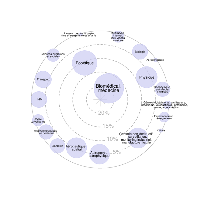
![]() Expertises du GdR - cartographie par mots-clés applicatifs - 19/09/2025