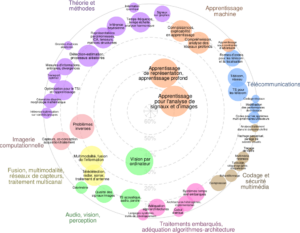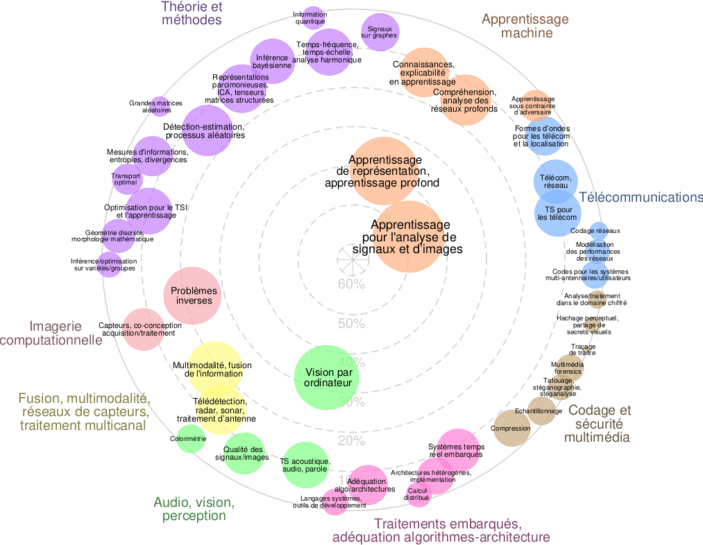
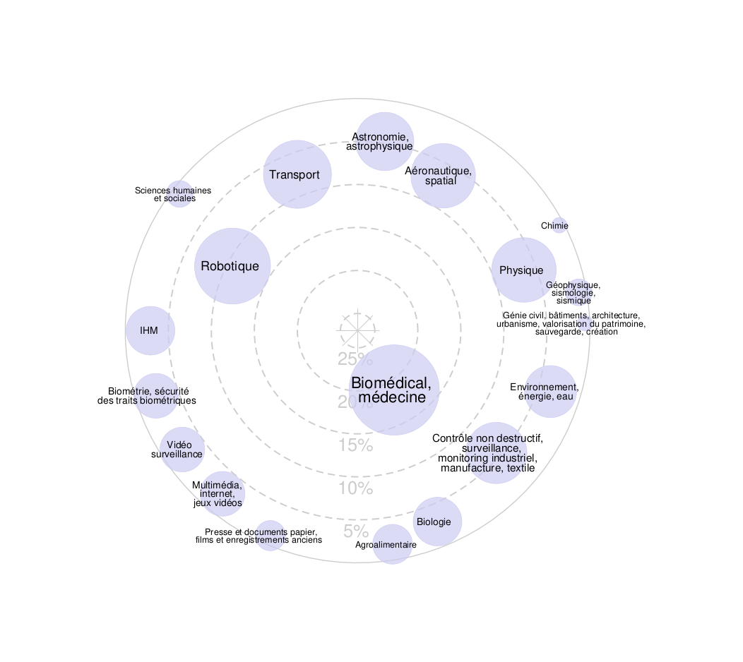
Assemblée Générale du GdR, 6-8 octobre 2025
La prochaine Assemblée Générale du GdR se déroulera à La Grande-Motte Presqu’Ile du Ponant, du...

28 Février 2024
Catégorie : Doctorant
PhD Advisors : Nicolas Verrier - Olivier HaeberlÉ
IRIMAS - IMTIS, 61, rue Albert Camus, 68 093 Mulhouse
Phone:+33 3 89 33 76 66/61 e-mail : nicolas.verrier@uha.fr olivier.haeberle@uha.fR
Quantitative phase imaging (QPI) becomes more and more popular in biomedical imaging, especially in optical microscopy. Unlike other methods relying on fluorescence of contrast agents, incorporated into the sample, QPI extracts phase and amplitude directly from the optical field transmitted or reflected by the object, rendering sample labeling optional.
Within the IMTIS (Multimodal Imaging, Information and Signal Processing) team at IRIMAS (Institut de Recherche en Informatique, Mathématiques, Automatique et Signal), we have been developing, for about 15 years now, a generalization of QPI called Tomographic Diffractive Microscopy (TDM) [1-4]. By varying the object's illumination conditions, it is possible to obtain a 3D reconstruction of its complex refractive index (in absorption and refraction), with improved resolution compared to conventionnal QPI approaches [1,5,6].
These methods offer an interesting alternative to flurorescence microscopy, but suffer from a lack of chemical selectivity in the reconstructed information. Indeed, very different structures may have a similar refractive index. The aim of this innovative PhD proposal is to develop new approaches, in order to restore selectivity to tomographic images.
Preliminary studies, illustrated in Figure 1, have shown that it is indeed possible to access quantitative polarization information in TDM, offering a structural selectivity to tomographic content, by distinguishing birefringent and non-
birefringent elements within the same specimen
[7,8]. Another possible approach is based on multispectral or hyperspectral imaging (spectro- imaging). In particular, it has already been proven taht the variation of absorption with wavelength enables chemical species to be distinguished at the micrometric scale. [9]. Finally, a dynamic selectivity is also possible: temporal heterodyning of the detected signal can be used to isolate moving structures within a sample by measuring the Doppler effect they induce onto the scatterd light [10].
The proposed work is therefore twofold: an
experimental aspect, to complete the
instrumental configurations needed to acquire the necessary data [5,6], and a numerical aspect, to improve tomographic image reconstruction algorithms using this new data [11]. New reconstruction/display modalities are also possible [12].
The successful candidate will have a sound knowledge of imaging and signal processing, and be proficient in an object-oriented programming language (C++, Python, Matlab, etc.). A taste for experimental work will also be highly appreciated. You will be part of a dynamic team with recognized scientific expertise, and benefit from its already available equipment and operating resources (to attend conferences, pay publication fees, etc.).
1. V. Georges, et al., Opt. Lett. 34, p. 79 (2009)
2. B. Simon, et al., J. Biophoton. 3, p. 462 (2010)
3. H. Liu, et al., Appl. Opt. 53, p. 748 (2014)
4. B. Simon, et al., Optica 4, p. 460 (2017)
5. E. Wolf, Opt. Comm. 1, 153 (1969)
6. V. Lauer, J. of Microscopy 205, 165 (2002)
7. A. M. Taddese et al., Opt. Express 31, 9034 (2023)
8. N. Verrier et al., J. Microscop. 289, 128 (2023)
9. Y. Sung, Phys. Rev. Appl., 10, 054041 (2018)
10. N. Verrier, et al., Opt. Express 22, 9368 (2014)
11. F. Yang, et al., Opt. Express 28, 3905 (2020)
12. R. Abbessi, et al., J. Microsc. 288, 193 (2022)